Wize High School Grade 11 Biology Textbook > Genetics of Inheritance
The Cell Cycle & Mitosis

0:00 / 0:00
Phases of The Cell Cycle
The central feature of life is that organisms grow, reproduce and die. Just like humans go through different stages in our growth and development, so do each of our cells.
- The cell cycle is like the lifecycle of a cell – from the beginning of a new cell, through growth, and finally division.
- Some cells are capable of dividing many times, while others are not.
- In order to divide, a cell must go through its "stages of life". In this time, the following things will happen:
- The cell will grow;
- It will replicate its DNA;
- It will produce new organelles;
- As it nears cell division, it will sort the DNA so that each daughter cell receives one copy of each chromosome;
- Split into two cells (twin daughter cells).
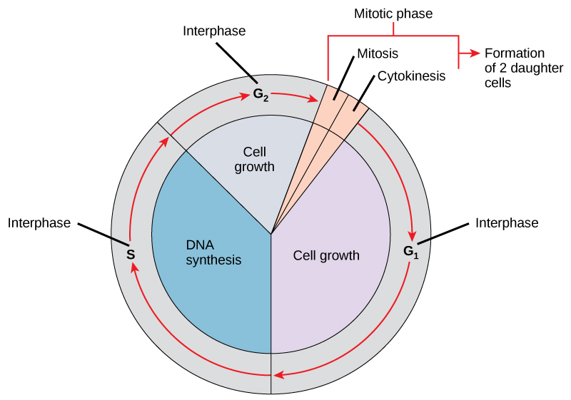
The cell cycle can be divided into 2 major phases:
- Interphase: Cell growth and DNA replication.
- G1 phase (1st gap)
- S phase (Synthesis)
- G2 phase (2nd gap)
- Mitotic (M) Phase: Replicated DNA and cell contents are separated.
- Mitosis
- Cytokinesis (partitioning of the cytoplasm)
Wize Tip
Think about it like this: cells can only think about reproduction! The cell is either waiting and preparing to divide (interphase), or is dividing (dividing).
Interphase
Interphase can be split into three separate phases:
- G1 phase: Gap phase 1.
- The cell is stocking up on nucleotides, proteins and energy to replicate its DNA. That is, it increases abundance of molecules involved in replication.
- S phase: Synthesis phase.
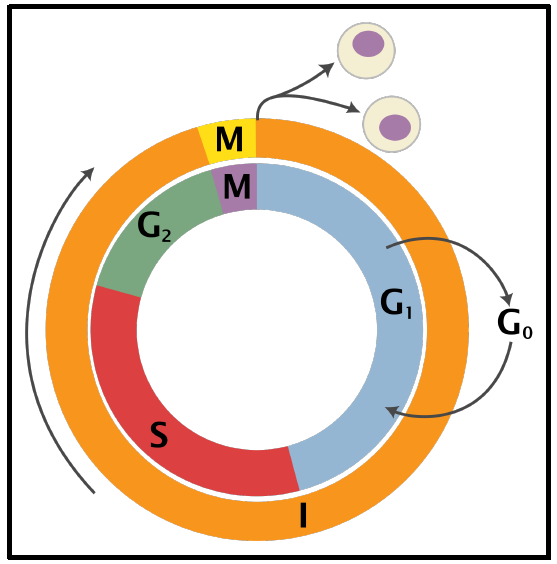
- The cell replicates its DNA to form sister chromatids, and replicates the microtubules organizing center called the centrosome.
- DNA remains in semi-condensed chromatin form.
- G2 phase: Gap phase 2.
- The cell may grow in size and increases abundance of proteins involved in mitosis (i.e. chromosome movement and manipulation).
- It also replenishes energy stores and some organelles are duplicated.
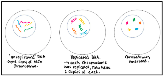
- There may also be a G0 phase, in which the cells exist in a quiescent state (quiet) and they are not in the process of dividing or preparing to divide. This can sometimes be seen as an extended G1 phase.
- May be temporary (e.g. due to lack of nutrients);
- May be permanent (e.g. adult heart cells usually do not divide).

0:00 / 0:00
Mitotic Phase
Mitosis (M Phase) is the part of the cell cycle in which a cell divides into two identical daughter cells, which are genetically identical to each other as well as the "mother" cell.
- Recall that the M Phase of the cell cycle can be subdivided into mitosis and cytokinesis.
- The first portion is also called karyokinesis, which literally just means division of the nuclear contents.
- The second portion, cytokinesis, means separation of the cytoplasmic components into two daughter cells.
- Most of the cell division in our bodies occurs through mitosis.
- Mitosis is for growth, and to replace old and dying cells.
Wize Tip
Remember that "-kinesis" just means motion.
Phases of Mitosis
- Prophase
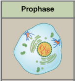
- Chromosomes start to condense.
- Centrosome begins to form the mitotic spindle structure composed of microtubules which organize chromosomes and move them to the appropriate locations during mitosis.
- The spindle gets longer as the centrosomes move to opposite poles of the cell.
- The nucleolus disappears and nuclear envelope breaks down.
- Prometaphase
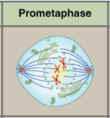
- Chromosomes continue to further condense.
- The mitotic spindle grows and begin to attach to the kinetochore of the chromosomes' centromere.
- The kinetochore is a protein located at the centromere of the sister chromatids.
- Metaphase
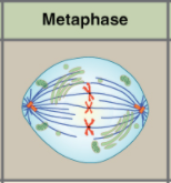
- Chromosomes align at the metaphase plate (also sometimes called the equatorial plane).
- The two kinetochores of each chromosome (one for each sister chromatid) are attached to microtubules – each attached to an opposite centrosome.
- Maximal condensation of chromosomes, but chromatids are still attached to one another.
Wize Tip
Chromatids align like the stars in order for the cell to divide... that's so meta!
- Anaphase
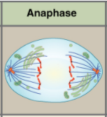
- Sister chromatids separate and are pulled towards opposite ends of the cell towards the two centrosomes.
- Now they are each one chromosome!
- The microtubules that are not attached to chromosomes push apart, causing the cell to elongate and the poles to move away from each other.
- Telophase
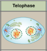
- The mitotic spindle is broken down into tubulin monomers that will form the cytoskeleton of daughter cells.
- Chromosomes start to decondense.
- Nuclear envelope begins to form.
Wize Tip
Cells are telling each other good bye!

0:00 / 0:00
Cytokinesis
This is sometimes seen as the second stage of M phase. Is very different for animals and plants.
- Physical separation of cytoplasmic components occurs in this phase.
- Usually starts to occur in late anaphase or telophase.
In animal cells
- The cell "pinches in" at the metaphase plate due to a band of filaments that act like a drawstring. These filaments are composed of a protein called actin.
- The pinching forms a crease called the cleavage furrow.
- Two daughter cells are formed when cytokinesis is complete.
In plant cells
- Remember that plants have cell walls, composed of cellulose, so a new one must form between the daughter cells.
- Vesicles formed from the Golgi apparatus that contain the components necessary for this process (e.g. glucose) go to the metaphase plate.
- A cell plate forms down the middle, splitting the cell into two and forming a cell wall between them.

0:00 / 0:00
Example: Mitosis & Chromosomes
Describe the phases of mitosis with respect to the chromosomes:
a. Prophase – duplicated chromosomes, as sister chromatids, condense, cohesins are interacting with sister chromatids
b. Prometaphase – spindle attachment to centromeres
c. Metaphase – spindle fibers pull sister chromatids to the centre of the cell, at the metaphase plate
d. Anaphase – cohesins no longer interact with sister chromatids, microtubule motor proteins move sister chromatids to opposing poles of the cell
e. Telophase – sister chromatids, now chromosomes, decondense
Practice: Cell Division Phase
Cells prepare for division in the ___ phase.
Practice: Lack of Actin
A human cell has received a mutation and is unable to produce actin. What would be the result after mitosis?
Practice: Chromosomes and Chromatid Number
If a cell contained 4 chromosomes during prophase, how many chromosomes and chromatids would it have during prometaphase?