Popular Courses
Find My CourseCytoskeleton
The cytoskeleton is a network of filaments that extend throughout a cell's cytoplasm. This network plays a major role in organizing the structures and activities of a cell and can be found in both eukaryotic and prokaryotic cells, with some notable differences.

The eukaryotic cytoskeleton is composed of three types of protein filaments: microtubules, intermediate filaments, and microfilaments (actin filaments).
Microtubules: Structure
- Made of globular proteins called tubulin:
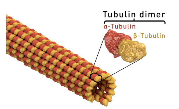
- Each tubulin protein is made of two slightly different subunits:
- Alpha- tubulin
- Beta-tubulin
- These two subunits will join together to form a dimer:
- Dimers will join together to form a long chain, that will arrange themselves into a sheet like structure and eventually roll up to form a hallow tube that is approximately 25nm in diameter.
Have structural directionality: Have two structurally distinct ends.
- One end will be anchored to the microtubule organizing center (MTOC). The other end, known as the plus end is where dimers can be quickly added or removed as needed, this is what makes microtubles dynamic structures.
- There are 2 different types of MTOC; centrosomes and basal bodies. Each having a similar structure but different functions.
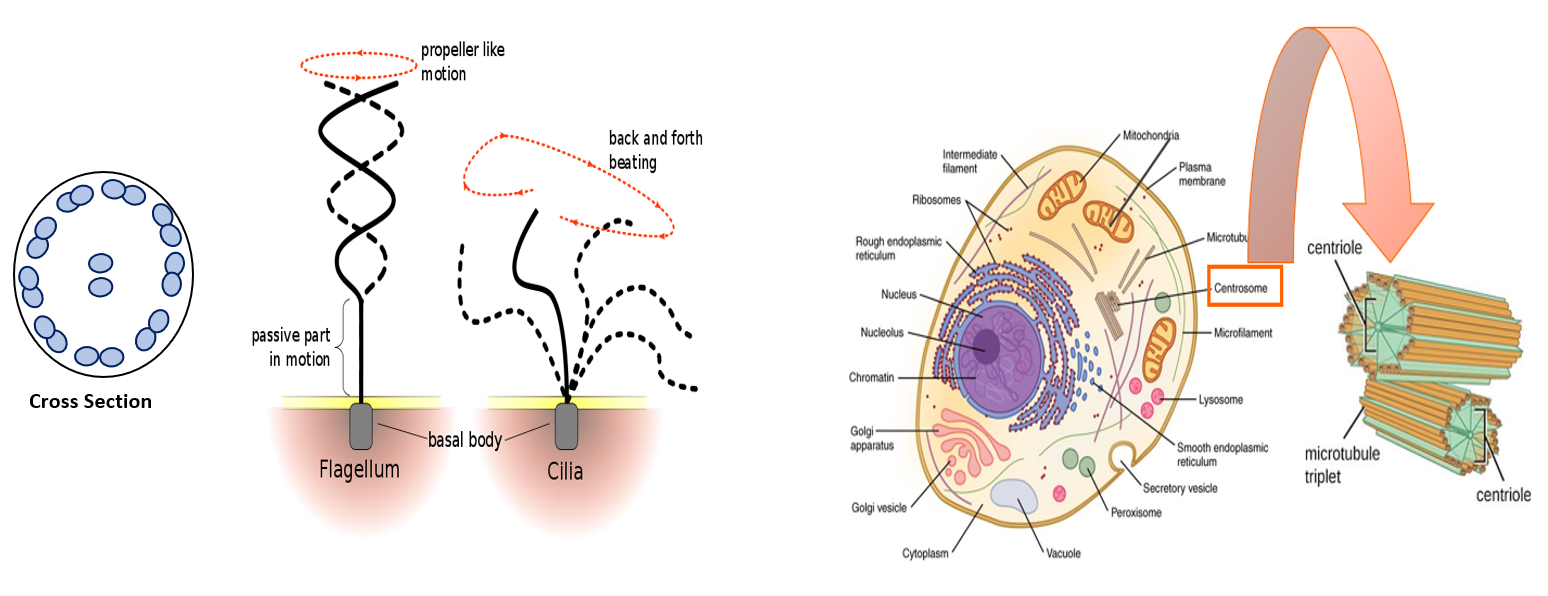
- Centrosome: In animal cells, microtubules grow from the centrosome, a structure near the nucleus. Within the centrosome is a pair of centrioles at right angles to each another, each consisting of 9 sets of three microtubules arranged in a ring. (Plant cells do have centrosomes, but they are simpler)
- Basal body: Organizes and anchors the microtubules of specialized eukaryotic structures: cilium (short, hair-like projections) and flagellum (long, tail-like projections). Each consisting of 9 sets of two microtubules with two single microtubules in its center. Bending of cilia or flagella is enabled by motor proteins.
Microtubule Function:
- Maintenance of cell shape and structure by resisting compression
- Direct organelle movement as well as transport vesicles to their site of deposition.
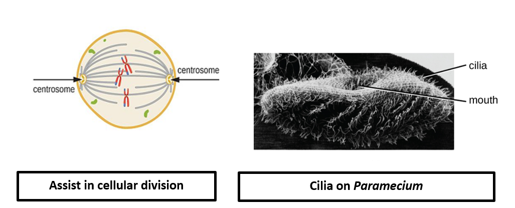
- Role in cell division [centrioles of the centrosome]
- Cell motility, propelling fluids across cells, and acquiring food [cilia and flagella]
- Example (cell motility): Human sperm cells have a flagellum so that it can swim to reach the oocyte.
- Example (propel fluids): The ciliated cells that line our respiratory tract help move microbes and debris up and out of the airways.
- Example (acquire food): The feeding current generated by Paramecium in its oral groove.
Intermediate Filaments: Structure
- Found in some animal cells
- Diverse class of cytoskeleton elements:
- Each type of intermediate filament is made up of a different type of fibrous protein.
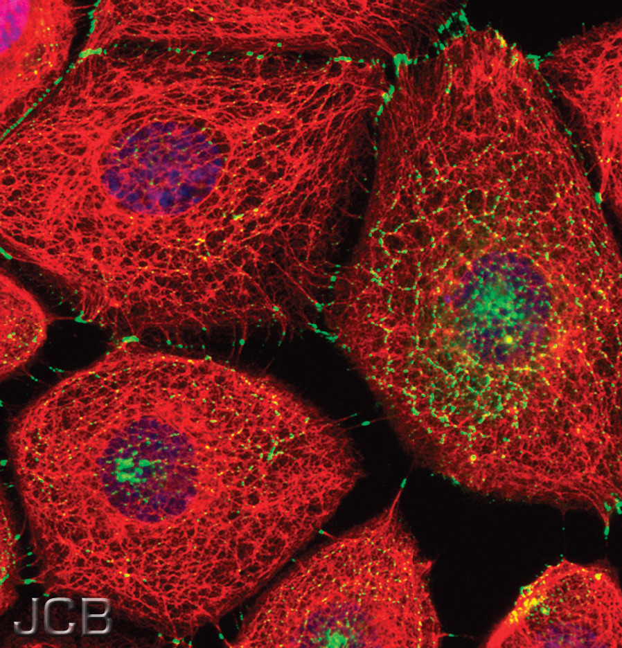
- Keratin proteins make up the two largest classes of intermediate filament proteins
- Fluorescent microscopy of keratin microfilaments is shown above.
- These proteins will join to form polymers, that will twist together to form a filaments that are approximately 10nm in diameter.
- Intermediate filaments have a "coiled cable" appearance
- Permanent structures
- They do not readily grow and disassemble; they are not dynamic
Intermediate Filaments: Functions
- Maintenance of cell shape and structure by resisting tension
- Anchorage of certain organelles, such as the nucleus
- Form the nuclear lamina
- Link cells together by forming special cell-to cell junctions
Microfilaments: Structure
- Made of globular proteins called actin:
- Many actin molecules join together to form actin polymers. When two actin polymers twist around one another they form a thin solid tube of approximately 7nm in diameter.
- Microfilaments can also be called actin filaments since they are made from actin monomers (building blocks)
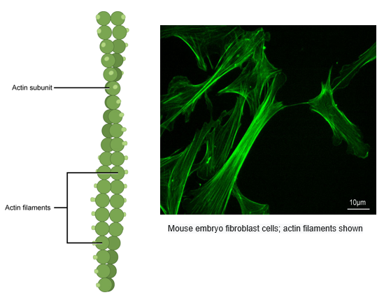
- Have structural directionality: Have two structurally distinct ends.
- Dynamic structures
- They can lengthen or shorten as needed.
Microfilaments: Function

- Maintenance of cell shape and structure by resisting tension
- Cellular events requiring motion
- Serve as "tracks" for the motor protein myosin, together they enable:
- Animal cells to pinch into 2 daughter cells during the final stage of cell division in which the cell physically separates (cytokinesis).
- Muscle contraction.
- The movement of transport vesicles and some organelles.
- Cell Motility
- Crawling of white blood cells and amoeba (unicellular eukaryote) is brought about my actin filaments interacting with myosin.
- Amoeba move by forming cellular extensions called pseudopods, these are filled with cytoplasm and primarily consist of actin filaments.
- Cytoplasmic streaming
- Actin-protein interactions bring about the circular motion of the cytoplasm, this in turn enables the transport of nutrients, proteins, and organelles within cells.
- Usually observed in large plant cells.
Practice: Cytoskeleton Proteins
Which of the following cytoskeleton proteins is involved in splitting apart two cells during cellular division? [select all that apply]
Practice: Cytoskeleton Structures
Which cellular structure may be the underlying cause for the lack of motility of sperm cells?
Practice: Cytoskeleton Proteins
Answer each question by writing the correct term beneath it: