Wize University Biology Textbook > Cell Communication
Types of Cell Receptors [Internal Receptors, GPCRs, RTKs, Ion Channels]
Popular Courses
MCAT
General Course
Biology
General Course
General Biology
University Study Guides
BIOL 102
Queen's University
Grade 12 Biology
Canada High School
DAT
General Course
General Biology
University Study Guides
BIOL 130
University of Waterloo
SC/BIOL 1000 3.00
York University
BIO 1141
University of Ottawa
BLG 143
Toronto Metropolitan University
BIOL 1010
Dalhousie University
BIO230H1
University of Toronto
BIOL 1201
Western University
BIOL 1P91
Brock University
BS 161
Michigan State University
BIOL 331
University of Calgary
BIOL 1020
University of Manitoba
BIOA01H3
University of Toronto
BISC 102
Simon Fraser University

0:00 / 0:00
Internal Receptors
Signal detection usually occurs through internal receptors or surface receptors (more common). Receptors are proteins located inside or on the cell surface which bind to the signaling molecule and transmit the signal to the cell's interior. There are several different types of cell receptors.
Internal Receptors
- Located on the cell's cytoplasm.
- Respond to hydrophobic ligands. Example: certain hormones in the human body.
- Often the receptor-ligand complex moves into the nucleus to regulate gene expression.

The following are cell-surface receptors. Remember that these are integral (transmembrane) proteins anchored on the cell's lipid bilayer. They perform signal transduction, or the relaying of information from the cell's exterior to the interior.

0:00 / 0:00
G-Protein Coupled Receptors (GPCRs)
The following are cell-surface receptors. Remember that these are integral (transmembrane) proteins anchored on the cell's lipid bilayer. They perform signal transduction, or the relaying of information from the cell's exterior to the interior.
GPCRs are a type of receptor which interacts with G proteins. Humans have over 1000 different types of GPCRs and they carry out a very wide array of functions in our cells.
- A G protein is attached to the intracellular side of the membrane.
- They have three subunits: the alpha, beta, and gamma subunits.
- G proteins have the ability to bind GTP and GDP.
- The variety of GPCRs comes from the variety of molecules they activate.
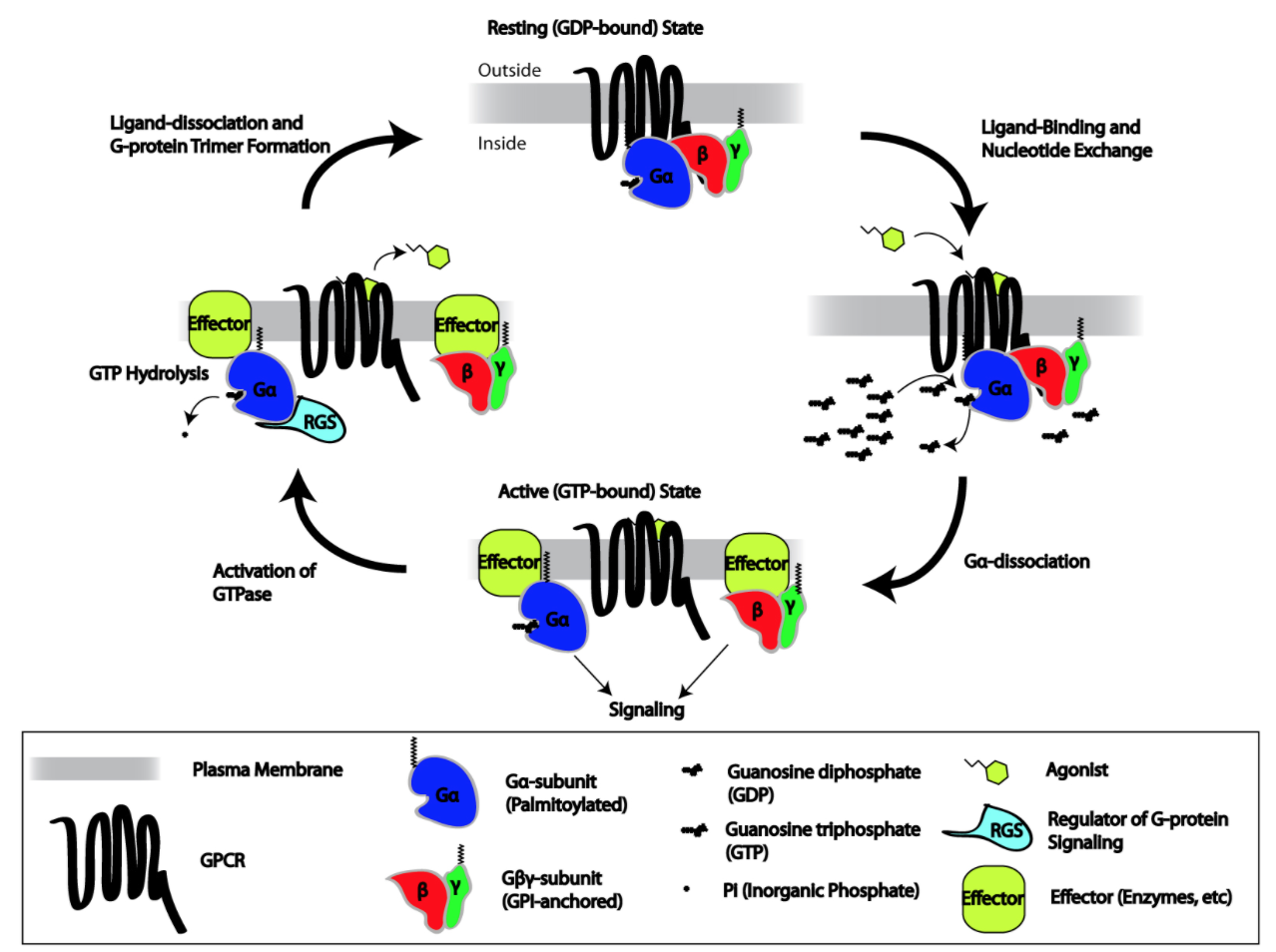
General GPCR pathway:
- Before the ligand binds, the G-protein is associated with its subunits, holding a GDP.
- The signal molecule binds to the cell surface receptor.
- Upon binding, the receptor undergoes a conformational change that activates the G-protein.
- The G protein's alpha subunit then lets go of GDP and picks up GTP and is now active.
- The G protein then dissociates from the GPCR and into alpha and beta-gamma subunits.
- These subunits can go activate other proteins as a result.
- Eventually the GTP is hydrolyzed to GDP.
- The subunits re-associate and the cycle starts again.
0:00 / 0:00
Receptor Tyrosine Kinases (RTKs)
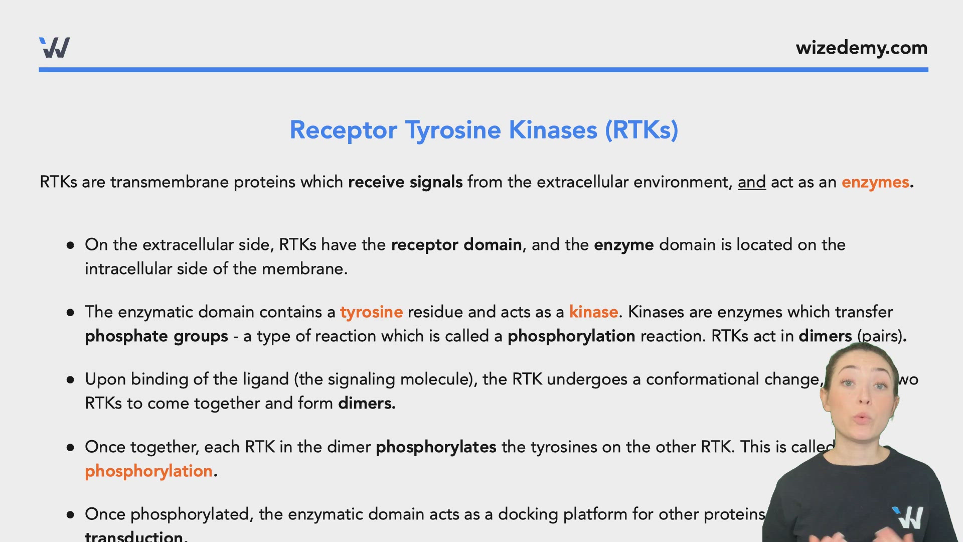
RTKs are transmembrane proteins which receive signals from the extracellular environment, and act as an enzymes.
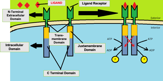
- On the extracellular side, RTKs have the receptor domain, and the enzyme domain is located on the intracellular side of the membrane.
- The enzymatic domain contains a tyrosine residue and acts as a kinase. Kinases are enzymes which transfer phosphate groups - a type of reaction which is called a phosphorylation reaction. RTKs act in dimers (pairs).
- Upon binding of the ligand (the signaling molecule), the RTK undergoes a conformational change, causing two RTKs to come together and form dimers.
- Once together, each RTK in the dimer phosphorylates the tyrosines on the other RTK. This is called cross phosphorylation.
- Once phosphorylated, the enzymatic domain acts as a docking platform for other proteins involved in signal transduction.

0:00 / 0:00
Ion Channels Receptors
Ion receptor channels are ion channels that open and close upon binding of a signaling molecule.
- Upon binding of the ligand (signaling molecule), it causes the channel to open.
- This allows for the flow of ions (e.g. K+, Na+, Ca+, Cl-) from an area of high concentration to an are of low concentration.

Practice: What Type of Receptor?
A certain hormone finds its receptor on a particular cell. When it binds, the receptor opens, allowing Ca2+ to rush into the cell. This receptor must be:
Practice: GPCRs
What is true about GPCRs?
Practice: Receptor Tyrosine Kinases (RTKs)
Which is true about receptor tyrosine kinases (RTKs)?