Wize High School Grade 11 Biology Textbook > Human Physiology: The Digestive System
Anatomy of the Digestive System

0:00 / 0:00
The Mouth
- Digestion begins at the oral cavity (the mouth) where food is ingested.
- Mechanical digestion by the teeth increases the surface area of the food, allowing for increased exposure to digestive enzymes. This process is called mastication.
- Chemical digestion also occurs in the mouth due to the secretion of enzymes such as salivary amylase, which breaks down carbohydrates.
- The tongue, one of the strongest muscles in the body, pushes food around into a ball called a bolus, which passes to the pharynx.
- To prevent food from entering the trachea when we swallow, the epiglottis folds over the opening, ensuring the bolus enters the esophagus.

The Esophagus
- A ~24 cm long tube connecting the mouth to the stomach
- Circular and longitudinal muscles allow for peristalsis, which mediates the pushing of food through the digestive tract.
- At the end of the esophagus, the bolus reaches the lower esophageal sphincter, a ring of muscles that prevents stomach acid from entering the esophagus, through which food must be pushed to reach the stomach.


0:00 / 0:00
The stomach
- The stomach is a muscular pouch which provides mechanical digestion by churning the bolus
- Propulsion (gentle mixing), grinding (strong mixing) and retropulsion (released into duodenum)
- Gastric juices contain acid and digestive enzymes, providing chemical digestion
- Hydrochloric acid (HCl) is secreted into the stomach by parietal cells, giving a pH ~1 - 2.5. The acidic pH kills viruses and bacteria and helps with chemical digestion
- The parietal cells produce H+ and Cl- ions separately, to prevent the accumulation of HCl intracellularly. They produce these ions in response to the peptide hormone gastrin.
- Gastrin is produced by G cells in the stomach lining
- To prevent damage to the cells lining the stomach, goblet cells aka mucus neck cells secrete a thick mucus layer
- The enzyme pepsin is a protease – it breaks down proteins into smaller peptides.
- Pepsin is secreted as pepsinogen and is converted to pepsin upon exposure to HCl. This prevents the proteins of the stomach lining from being degraded. Pepsinogen is secreted by chief cells

Stomach Ulcers
- Caused by the bacterium Helicobacter pylori
- The bacterium burrows into the mucus layer to protect itself from the acidic pH, and then adheres to the epithelial cells beneath the mucus.
- To further protect itself from the acidic pH, the bacterium produced urease, which helps to neutralize the stomach
- Urease leads to the production of ammonia, which damages the epithelial cells.
- An immune response is triggered, leading to inflammation of the stomach lining, eventually leading to ulcer formation


0:00 / 0:00
The Small Intestine
- After being further digested in the stomach, the food is now called chyme, which is passed to the small intestine
- The small intestine is where the majority of nutrient absorption occurs
- It can be divided into three sections:
- The duodenum: location of bile duct.
- The jejunum: where the majority of the abosroption takes place.
- The ileum: involved in absorption and compaction of what is left over, passing it on to the large intestine.

Digestive enzymes and chemicals are secreted into the intestine to allow for chemical digestion of the chyme:
- Bile is produced by the liver and aids in chemical digestion by emulsifying the lipids, exposing them to digestive enzymes.
- The pancreas secretes HCO3- to neutralize the pH of the chyme
- The pancreas also produced digestive enzymes: pancreatic proteases (eg. Trypsin and chymotrypsin), pancreatic amylase (breaks down carbohydrates), and pancreatic lipase (breaks down fats) and nucleases (break down nucleic acid polymers)
- The intestinal glands produce digestive enzymes: maltase and protease (enterokinase, aminopeptidase, dipeptidase).
- The lining of the small intestine has tiny, finger-like projections called villi, which increase the surface area of the intestine for absorption of nutrients
- Each vilus itself has microvilli, further increasing surface area


0:00 / 0:00
The Large Intestine
- Once all of the nutrients have been absorbed, the colon is responsible for the absorption of any remaining water and minerals
- Water crosses the membranes of the cells lining the colon by osmosis (movement of water along its concentration gradient)
- The colon is home to many different species of bacteria
- Once the rectum is full of fiber and undigested material, the anal sphincter loosens and the waste can be excreted

Motility
- Gastroileal Reflex - the valve between the ileum of the small intestine and the cecum of the large intestine (ileocecal valve) opens when the stomach senses food has entered
- Haustal Churning - the Haustae are the large kinks int he body of the large intestine. They cause churning of the waste
- Some peristalsis also occurs
0:00 / 0:00
What are the types of digestion?
What type of digestion occurs in the
a) mouth
b) esophagus
In the mouth: Chemical (saliva) and mechanical (teeth) digestion
In the esophagus: none

0:00 / 0:00
Accessory organs of the GI Tract
3 main glands
- Pancreas
- Liver
- Gal bladder

https://pixabay.com/vectors/digestive-system-human-digestion-41529/
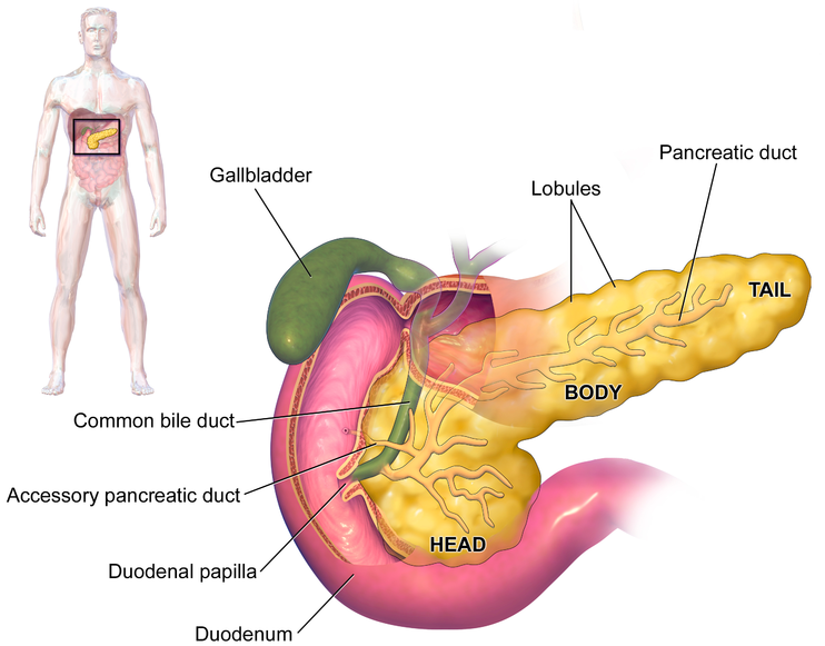
Pancreas
- long shaped gland that feeds hormones into the duodenum of the small intestine
- has a common duct with the gal bladder (bile duct)
- Exocrine secretions of the pancreas include
- Amylase
- Lipase
- Trypsinogen --> trypsin (activated by enteropeptidase from the brush border)
- chymotrypsinogen --> chymotrypsin
- procarboxypeptidase --> carboxypeptidase
- phospholipase
- colipase
- The endocrine hormones insulin and glucagon are secreted from the islets of Langerhans and are spread throughout the body.
https://commons.wikimedia.org/wiki/File:Blausen_0699_PancreasAnatomy2.png
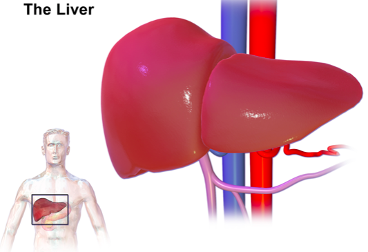
Liver
- made of liver cells called hepatocytes
- highly vascularized, sending and receiving many substances in the body.
- It receives a normal blood supply to keep it healthy (artery and vein for O2 and CO2 exchange
- it also has an other vein attached which comes from other parts of the body and stops by on its way to the heart. It can deliver things to liver from the body. This special vein is called the heptic portal vein.
- jack of all trades... has over 500 functions in the body
- for this class, we just need to know that it:
- secretes bile which is then stored in the gallbladder.
- excretes bilirubin (pigment) into the blood as waste
- stores and produces nutrients
- processes drugs and hormones
- gluconeogenesis (production of glucose from smaller molecules)
commons.wikimedia.org
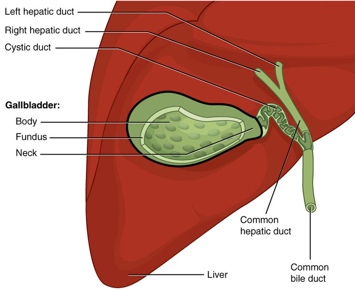
Gallbladder
- Stores bile
- bile is made of...
- bile salts
- cholesterol
- bilirubin
- water and ions
commons.wikimedia.org
0:00 / 0:00
List the main function of each digestive accessory structure in regards to the digestive tract
Liver: Bile production
Gallblader: Bile storage
Pancreas: produce the majority of enzymes needed to breath down chyme in the small intestine including....
- Amylase
- Lipase
- Trypsinogen --> trypsin (activated by enteropeptidase from the brush border)
- chymotrypsinogen --> chymotrypsin
- procarboxypeptidase --> carboxypeptidase
- phospholipase
- colipase
Practice: Digestive System
What moves food through the digestive tract?
Practice: Bile Production & Storage
Match the following organs with their function.
A.
Organ that stores bile
B.
Organ that produces digestive enzymes that work together with bile
C.
Organ that uses bile for digestion
D.
Organ that produces bile
Liver
Gallbladder
Small intestine
Pancreas
Practice: The Pancreas
Which of the following would occur if the pancreas stopped functioning?