Wize University Physiology Textbook > Sensory Physiology
Visual Modality
Popular Courses
MCAT
General Course
DAT
General Course
Intro to Physiology
University Study Guides
PHYSIOL 1021
Western University
PHYSIOL 2130
Western University
Intro to Physiology
University Study Guides
PSL300H1
University of Toronto
PHYSL 210
University of Alberta
PHGY 210
McGill University
PHGY 215
Queen's University
KNES 259
University of Calgary
PHGY 216
Queen's University
BIOL 273
University of Waterloo
BIOL 260
University of British Columbia
PHYSIOL 3120
Western University
KNES 260
University of Calgary
BIOL 116
Case Western Reserve University
PPT 301
University of North Dakota
ANAT 212
McGill University
HTHSCI 2FF3
McMaster University
0:00 / 0:00
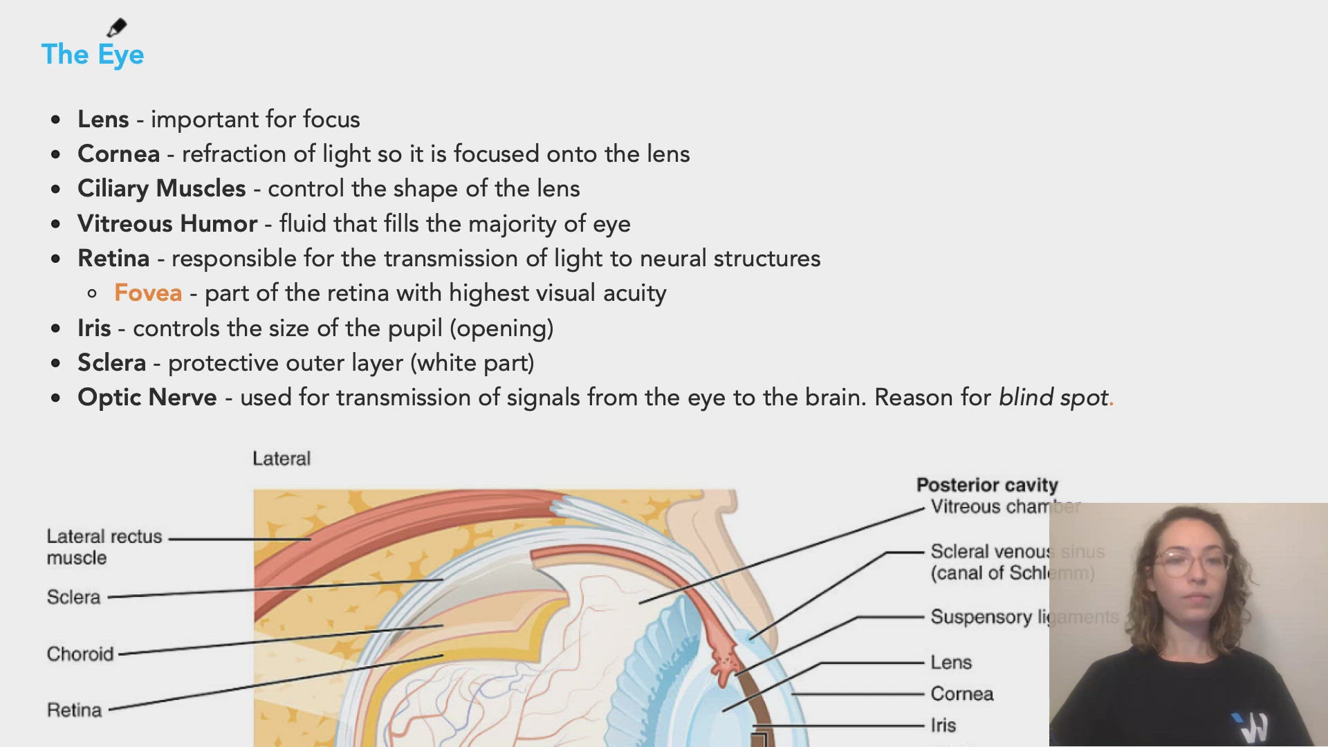
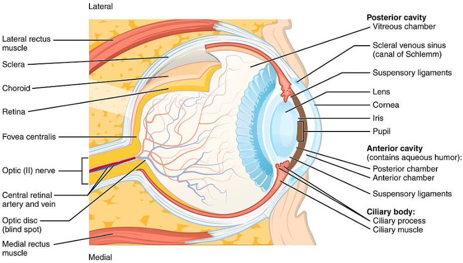
The Eye
- Lens - important for focus
- Cornea - refraction of light so it is focused onto the lens
- Ciliary Muscles - control the shape of the lens
- Vitreous Humor - fluid that fills the majority of eye
- Retina - responsible for the transmission of light to neural structures
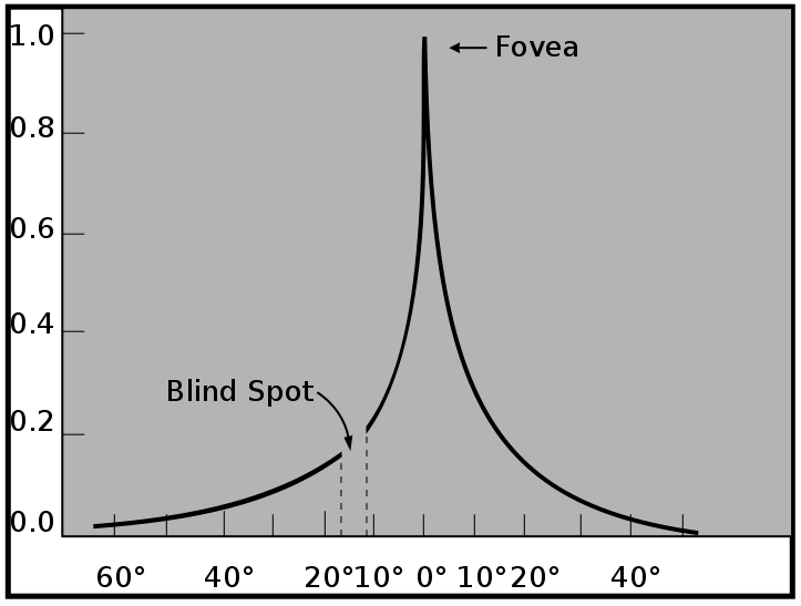
- Fovea - part of the retina with highest visual acuity
- Iris - controls the size of the pupil (opening)
- Sclera - protective outer layer (white part)
- Optic Nerve - used for transmission of signals from the eye to the brain. Reason for blind spot.
commons.wikimedia.org
commons.wikimedia.org
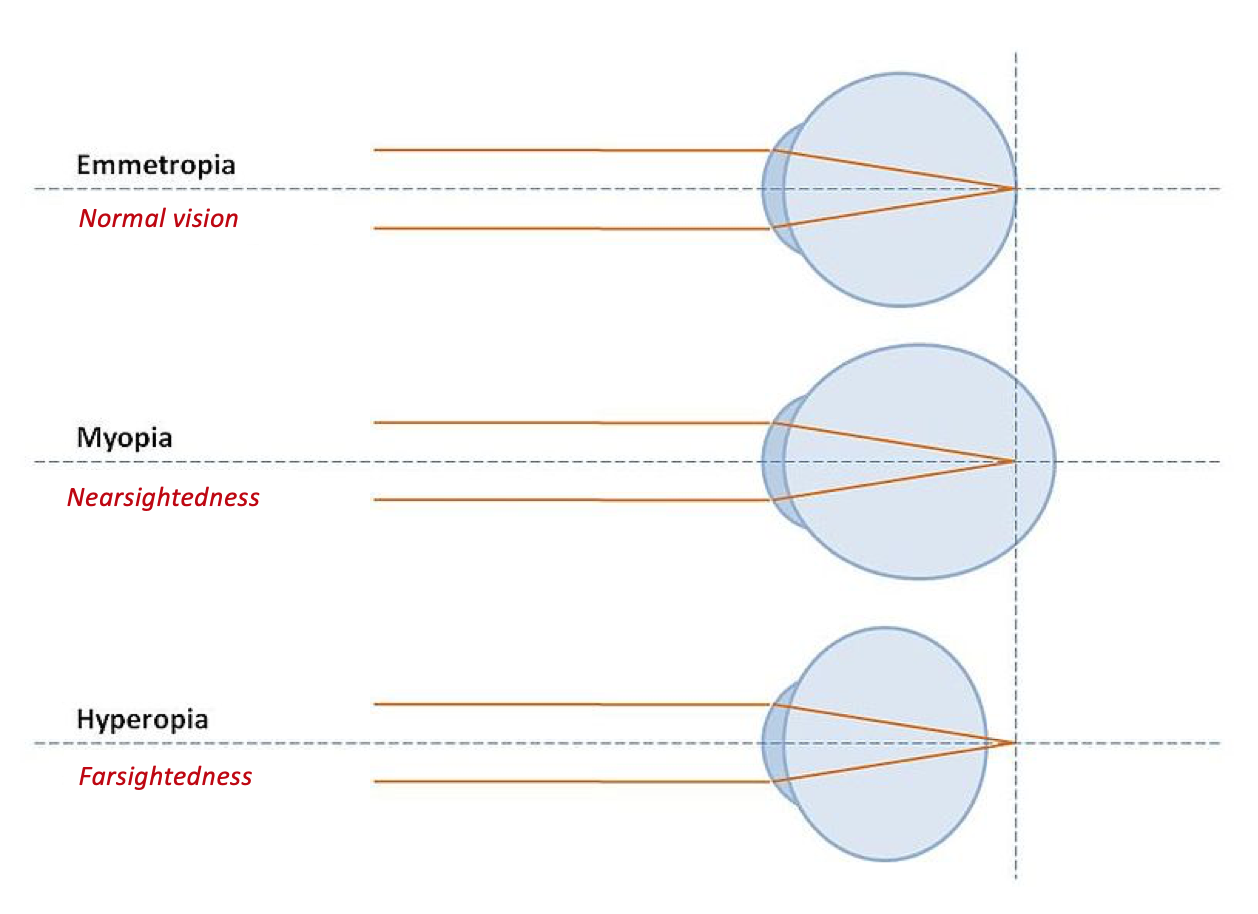
Common Eye Problems
- Myopia (nearsightedness) - eyeball too long
- Hyperopia (farsightedness) - eyeball too short
0:00 / 0:00
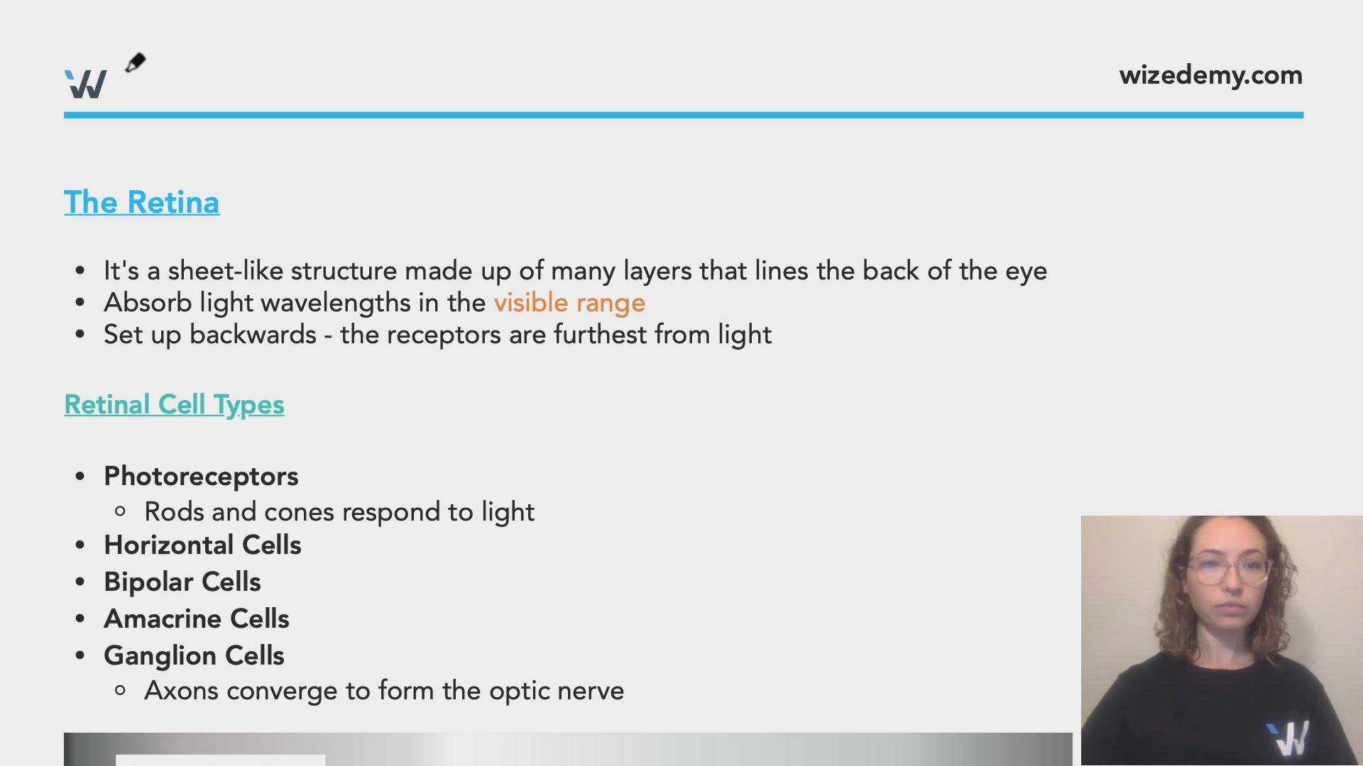
The Retina
- It's a sheet-like structure made up of many layers that lines the back of the eye
- Absorb light wavelengths in the visible range
- Set up backwards - the receptors are furthest from light
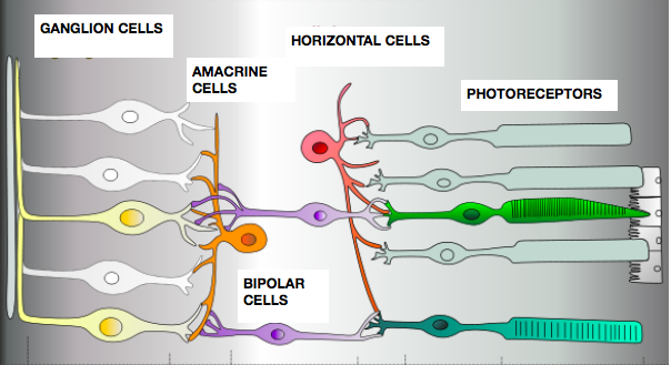
Retinal Cell Types
- Photoreceptors
- Rods and cones respond to light
- Horizontal Cells
- Bipolar Cells
- Amacrine Cells
- Ganglion Cells
- Axons converge to form the optic nerve
commons.wikimedia.org
Photoreceptors
- Involved in phototransduction
- Rods
- Black and white vision
- Relied on in the dark
- Found in peripheral vision
- High sensitivity: single photon causes many Na+ channels to close
Light strikes opsin --> Protein cascade --> Closing of Sodium Channels --> Hyperpolarization
- Cones
- Color vision (Cones are for Color)
- Trichromatic - three different types of cones that each respond to one of the primary colors
- Relied on during the day
- Found in high amounts in central vision (fovea)
- Low sensitivity
- When transitioning between dark and light environments, there's an adaptation period that corresponds to switching between rods or cones becoming active or inactive
0:00 / 0:00
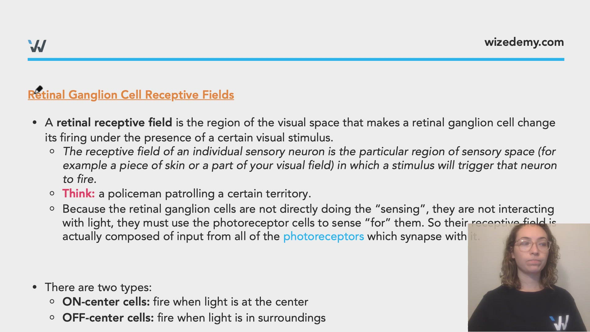
Retinal Ganglion Cell Receptive Fields
- A retinal receptive field is the region of the visual space that makes a retinal ganglion cell change its firing under the presence of a certain visual stimulus.
- The receptive field of an individual sensory neuron is the particular region of sensory space (for example a piece of skin or a part of your visual field) in which a stimulus will trigger that neuron to fire.
- Think: a policeman patrolling a certain territory.
- Because the retinal ganglion cells are not directly doing the “sensing”, they are not interacting with light, they must use the photoreceptor cells to sense “for” them. So their receptive field is actually composed of input from all of the photoreceptors which synapse with it.
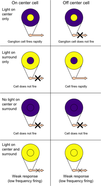
- There are two types:
- ON-center cells: fire when light is at the center
- OFF-center cells: fire when light is in surroundings
commons.wikimedia.org
0:00 / 0:00
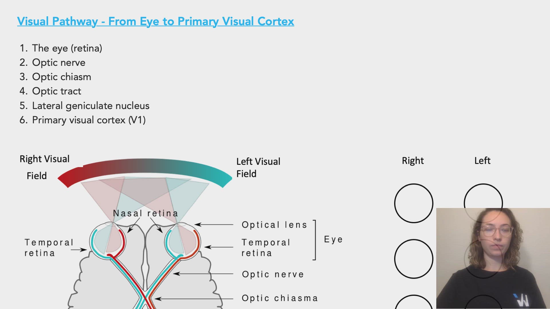
Visual Pathway - From Eye to Primary Visual Cortex
- The eye (retina)
- Optic nerve
- Optic chiasm
- Optic tract
- Lateral geniculate nucleus
- Primary visual cortex (V1)

Source: By Miquel Perello Nieto - Own work, CC BY-SA 4.0, https://commons.wikimedia.org/w/index.php?curid=37868501
0:00 / 0:00
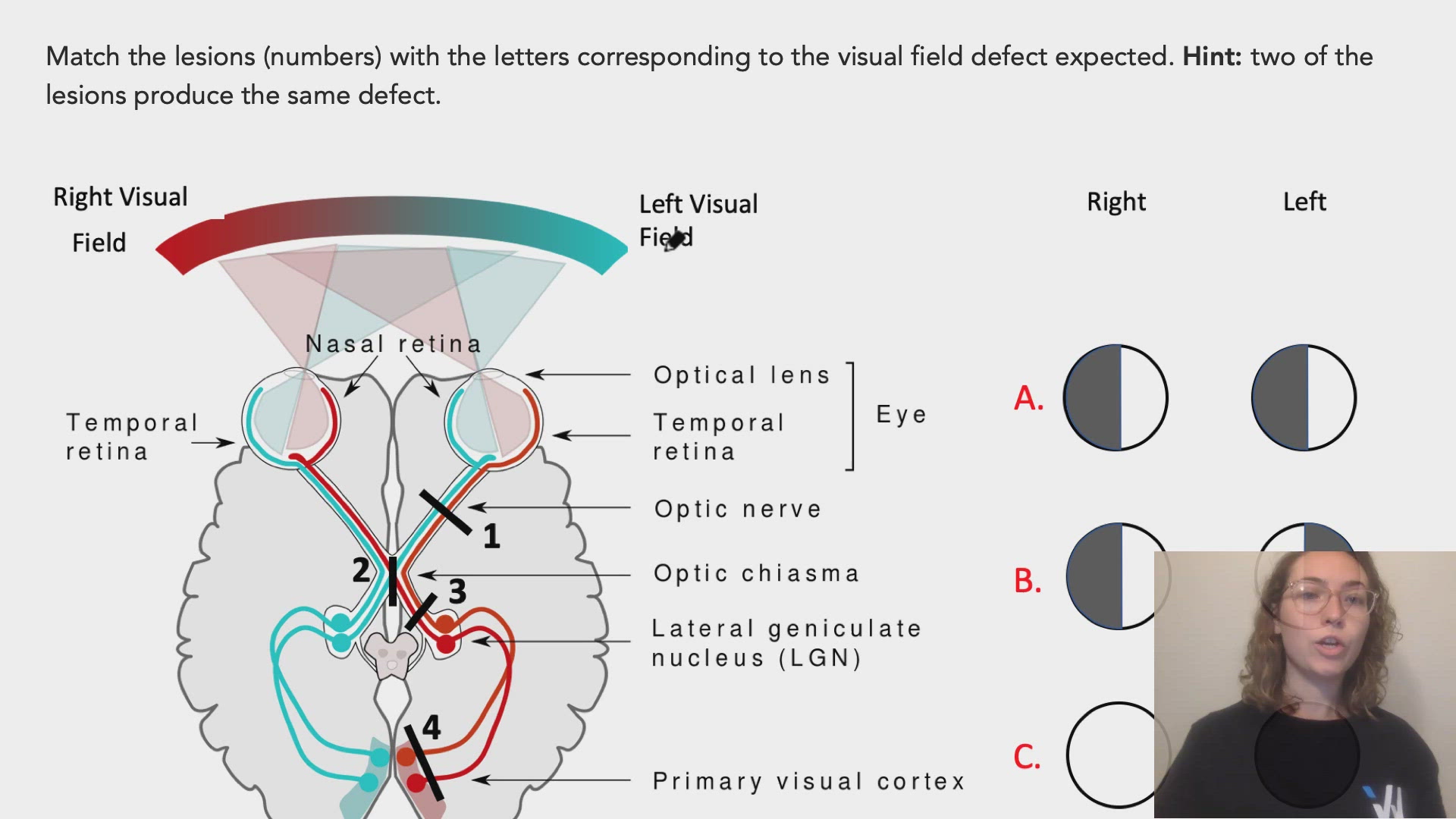
Match the lesions (numbers) with the letters corresponding to the visual field defect expected. Hint: two of the lesions produce the same defect.
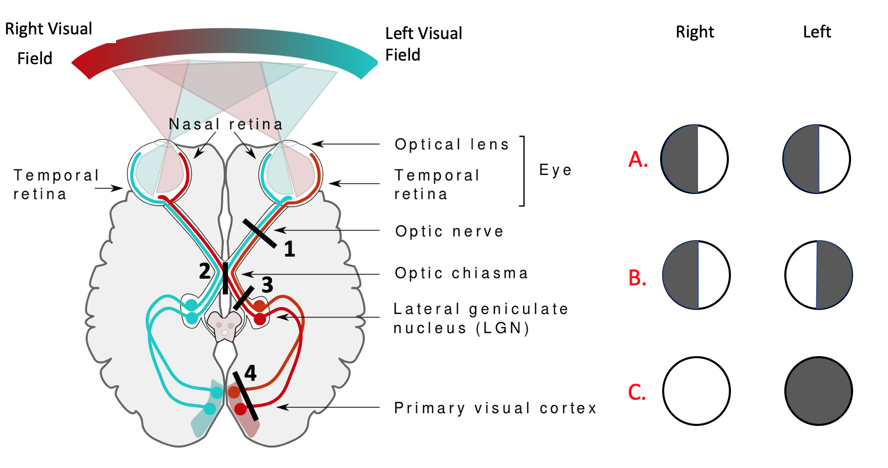
Lesion 1:
C
Lesion 2:
B
Lesion 3:
A
Lesion 4:
A
You and your cousin Mose are painting in the dark and someone suddenly turns on the light. You are temporarily blinded and it takes some time for your eyes to adjust to the new bright environment. Why does this occur?
What would cause loss of vision on the left hemifield of both eyes? (i.e. nasal visual field of the right eye and the temporal visual field of the left eye)