Wize University Physiology Textbook > Skeletal Muscle Physiology
Skeletal Muscle Physiology [more detailed]
Popular Courses
MCAT
General Course
DAT
General Course
Intro to Physiology
University Study Guides
PHYSIOL 1021
Western University
PHYSIOL 2130
Western University
Intro to Physiology
University Study Guides
PSL300H1
University of Toronto
PHYSL 210
University of Alberta
PHGY 210
McGill University
PHGY 215
Queen's University
KNES 259
University of Calgary
PHGY 216
Queen's University
BIOL 273
University of Waterloo
BIOL 260
University of British Columbia
PHYSIOL 3120
Western University
KNES 260
University of Calgary
BIOL 116
Case Western Reserve University
PPT 301
University of North Dakota
ANAT 212
McGill University
HTHSCI 2FF3
McMaster University

0:00 / 0:00
Physiology of Muscle Contraction
Types of muscle:
- Cardiac muscle - heart contraction
- Smooth muscle - involuntary contraction such as in gut, uterus, blood vessels
- Skeletal muscle - stripped and with many nuclei
Structure of Skeletal Muscle:
- 1 muscle = several fiber bundles
- 1 bundle = several muscle fibers (cells)
- 1 muscle fiber = several myofibrils
- 1 myofibril has several actin & myosin filaments arranged in end-to-end sarcomeres


commons.wikipedia.org
Muscle Contraction
- Sliding-filament model = thick filaments (myosin) move past thin filaments (actin)
- Sarcomeres shorten while filaments remain same length
- Requires actin-myosin binding sites, ATP and

0:00 / 0:00
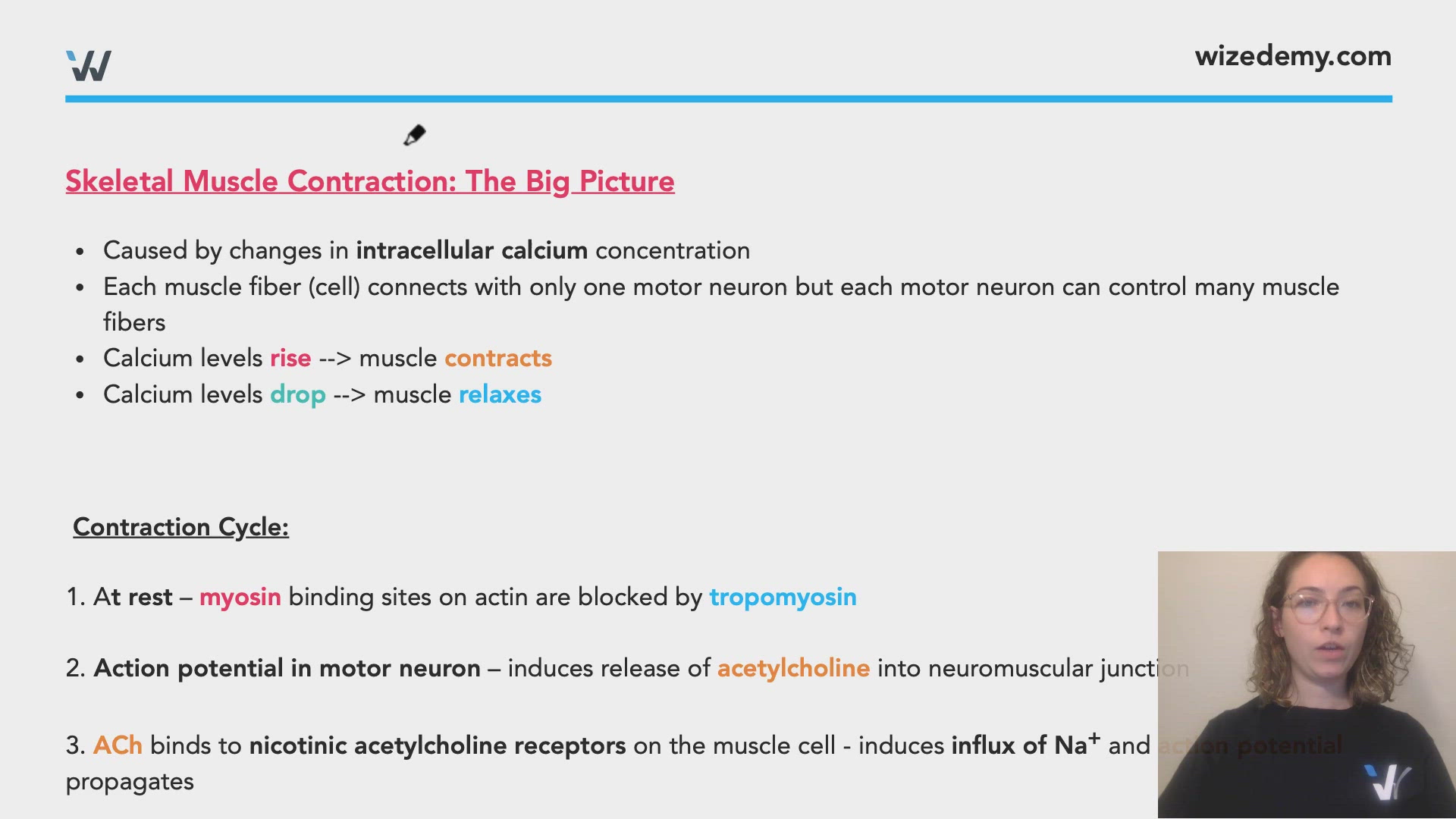
Skeletal Muscle Contraction: The Big Picture
- Caused by changes in intracellular calcium concentration
- Each muscle fiber (cell) connects with only one motor neuron but each motor neuron can control many muscle fibers
- Calcium levels rise --> muscle contracts
- Calcium levels drop --> muscle relaxes
Contraction Cycle:
1. At rest – myosin binding sites on actin are blocked by tropomyosin
2. Action potential in motor neuron – induces release of acetylcholine into neuromuscular junction
3. ACh binds to nicotinic acetylcholine receptors on the muscle cell - induces influx of Na+ and action potential propagates
4. Voltage-gated channels (called DHP receptor) open to allow calcium influx
5. This triggers the opening of Ryanodine receptors, allowing calcium to exit the sarcoplasmic reticulum into cytosol
6. Calcium binds troponin and pull off tropomyosin - myosin can bind to actin (cross-bridge formation)
7. Reset: pumped back into sarcoplasmic reticulum


commons.wikipedia.org
Cross-Bridge Cycle:


0:00 / 0:00
Length-Tension Relationship:
- There's an optimal amount of overlapping between actin and myosin heads to generate the most muscle tension (force generated by a muscle)
- Too much overlap --> not enough room for sliding
- Too little overlap --> not enough cross-bridges can form
- Summation: several action potential bursts add up to generate sustained contraction
- Recruitment: recruitment of additional motor neuron-muscle fiber pairs helps increase muscle tension
0:00 / 0:00
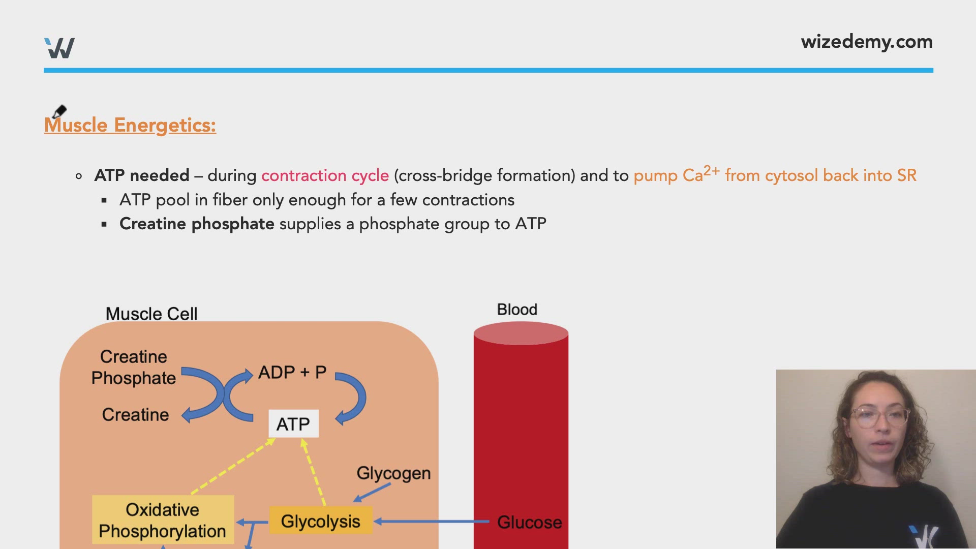
Muscle Energetics:
- ATP needed – during contraction cycle (cross-bridge formation) and to pump Ca2+ from cytosol back into SR
- ATP pool in fiber only enough for a few contractions
- Creatine phosphate supplies a phosphate group to ATP

Types of Skeletal Muscle Fibers:
- Classified by ATP source and speed of muscle contraction:
- Slow twitch fibers = contract slower but sustained longer, all oxidative
- Fast twitch fibers = rapid but shorter contractions, glycolytic or oxidative

Muscle Fatigue
- Protection from damage due to overuse
- Several factors come into play for this and is not fully understood
- Lactic acid buildup
- Changes in ion gradients
- CNS-mediated
0:00 / 0:00
You have the chance to visit a cadaver lab in order to see human organs up close. You notice that some of the cadavers have their muscles tensed up, which is called rigor mortis. Why does this occur?
Rigor mortis occurs because of the lack of ATP that ensues after death. ATP is required to break the cross-bridges between actin and myosin, which allows the muscles to relax. In the absence of ATP, the muscles remain in contraction.
Which of the following are true regarding skeletal muscle?
What is incorrect about different skeletal muscle fibers?
Select the correct statement: