Wize High School Grade 11 Biology Textbook > Cell Biology
Observing Cells Under the Microscope
0:00 / 0:00
Brightfield (Light) Microscopes
The invention of microscopes gave scientists a window into the world of microbes. Prior to these technologies, scientists could only hypothesize about how life was organized at the molecular level. As microscopy technologies have progressed, scientists have been able to learn more about how cells look and function.
Light microscopes were the first to be invented

- Light microscopes use glass lenses and light to magnify cells
- Allowed scientists to see the size, shape and contents of cells
- Led to the discovery that plants and animals have different cellular structures


Onion cells under a light microscope Cheek cells under a light microscope
There are three considerations when it comes to microscopy:
- Magnification: The image size produced by the microscope is much larger than the actual size of the object
- Resolution: Ability to distinguish between two adjacent objects
- Clarity: Ability to identify different structures
Light microscopes have 200 nm resolution and >2000x magnification.
Parts of a brightfield (light) microscope
- Ocular lens: the eyepiece lens. It usually magnifies 10x.
- Objective lens: there are usually 3 objective lenses on a microscope that magnify 4x, 10x, 40x (sometimes there is a 4th lens that magnifies 100x).
- Stage: platform the microscope slide is placed on.
- Slide holder: holds the slide to the platform.
- Condenser: focuses the light onto the specimen.
- Condenser aperture lever: controls how far the light condenser is from the slide. Not all microscopes have this.
- Light / Illuminator: produces the light that passes through the specimen.
- Coarse adjustment: this is always the larger of the two adjustment knobs.
- Fine adjustment: the smaller of the two adjustment knobs. Used to make smaller changes to the focus.
0:00 / 0:00
Electron Microscopes
Light microscopes do not have high enough resolution to allow scientists to pick out fine cellular details. Most cellular structures are too close together (less than 0.2 µm) for light microscopes to distinguish.
The invention of the electron microscope allowed scientists to understand cellular structures, such as the cell membrane, in much higher detail.
- Electron microscopes use beams of electrons instead of light to produce images.
- Compared to light, this beam has a very short wavelength, allowing it to pass between two cell features less than 0.2 µm apart.
The first usable electron microscope was built in 1938 at the University of Toronto by two graduate students, James Hillier and Albert Prebus.
Transmission Electron Microscopes (TEM)
- The beam of electrons is passed through a sample that has been sliced into very thin sections
- Produces a 2-dimensional image
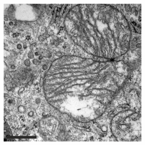
TEM of a mitochondria
Scanning Electron Microscopes (SEM)
- The beam of electrons is scans the surface of a sample
- Produces a 3-dimensional image
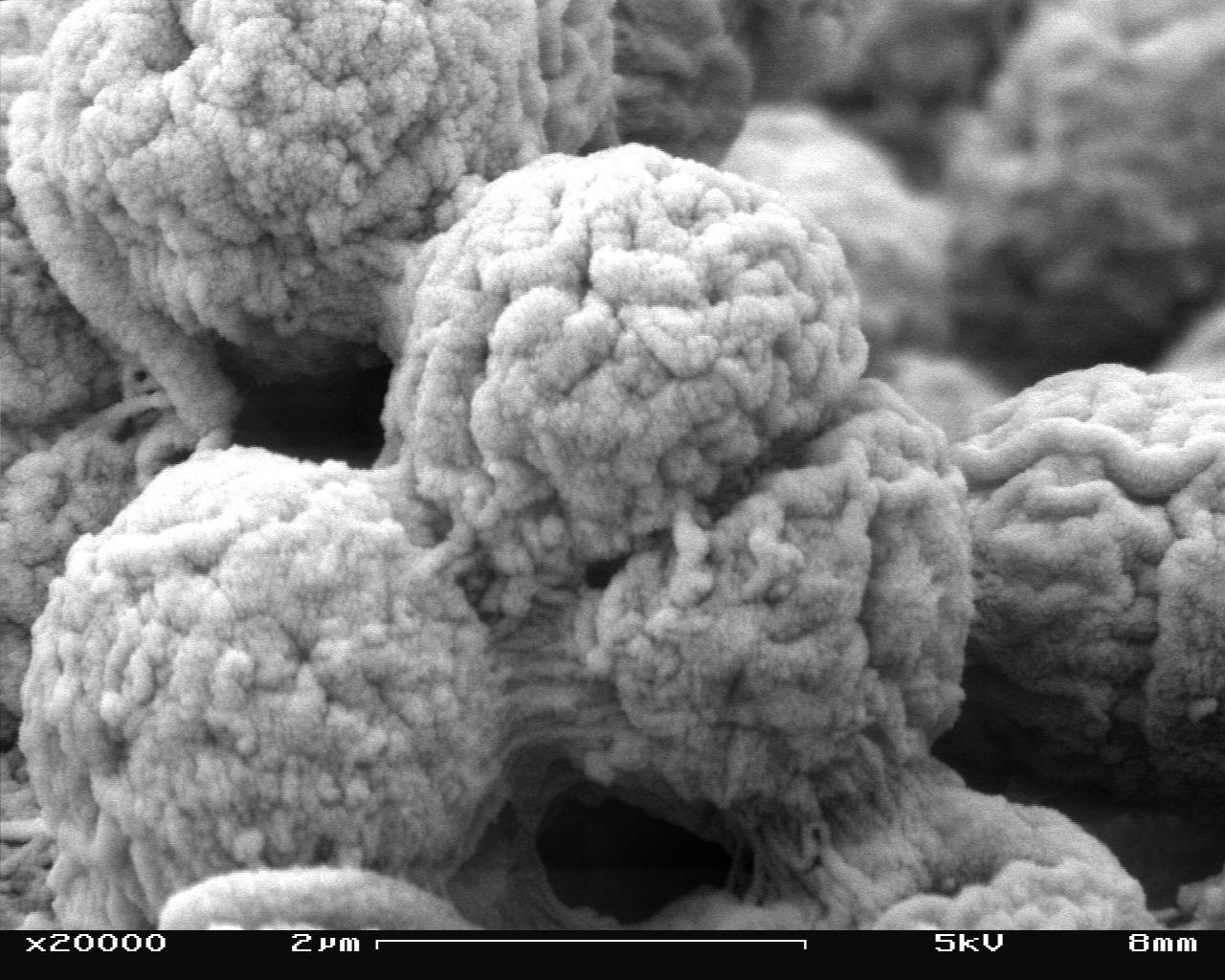
SEM of algae cells
Scanning Tunnelling Microscopes (STM)
- A tiny electrical probe is placed near the surface of the sample,
- Produces a topographical image
- Used for atomic-level imaging
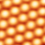
STM of silicone atoms
0:00 / 0:00
Determining Magnification & Size
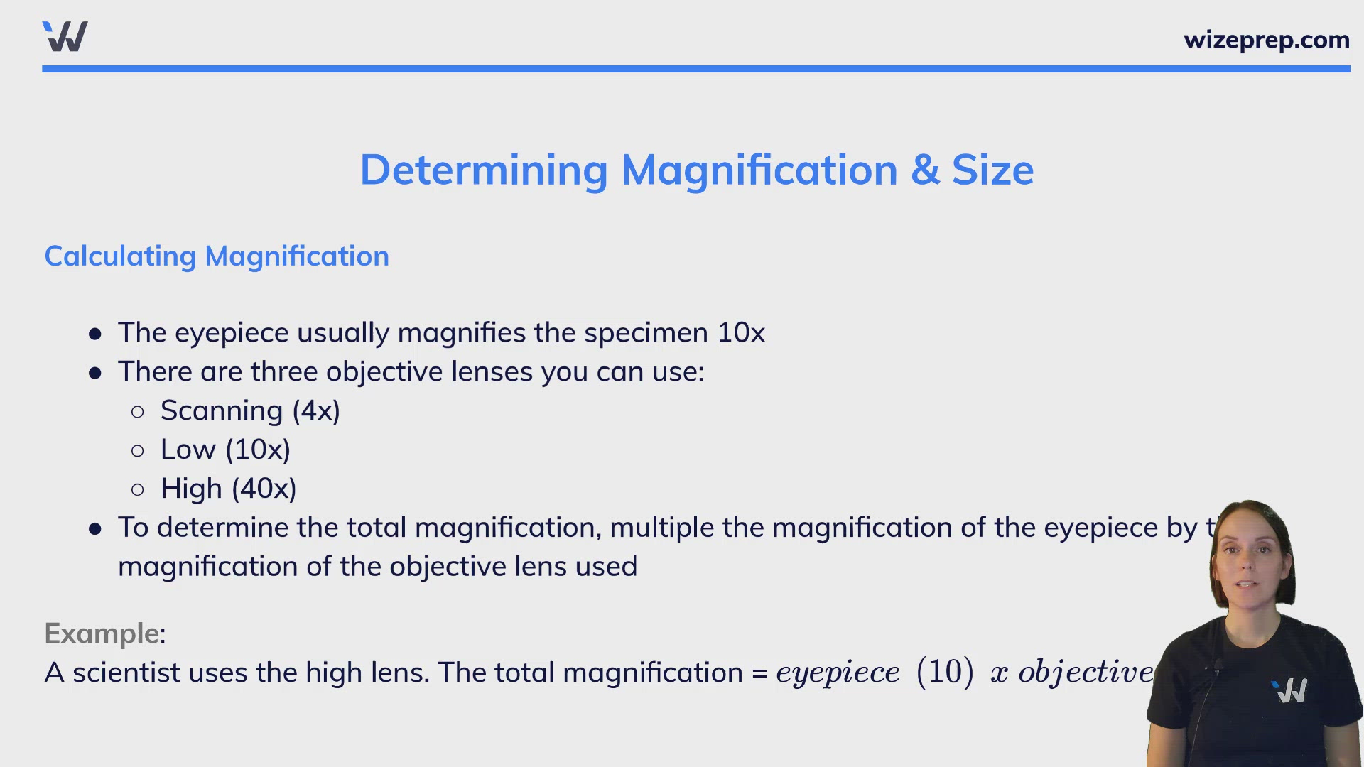
Calculating Magnification
- The eyepiece usually magnifies the specimen 10x
- There are three objective lenses you can use:
- Scanning (4x)
- Low (10x)
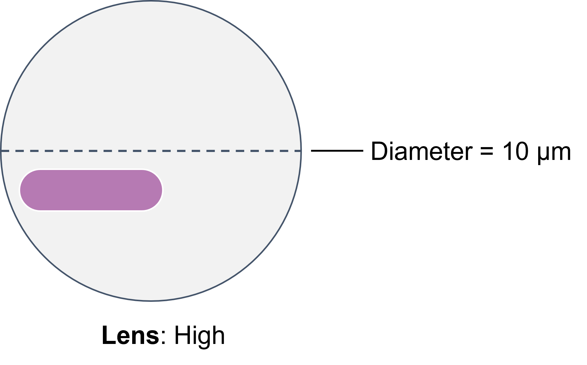
- High (40x)
- To determine the total magnification, multiple the magnification of the eyepiece by the magnification of the objective lens used
Example:
A scientist uses the high lens. The total magnification =
Estimating Size
To estimate the size of a specimen, the field of view must first be determined. This can be found by focusing on a transparent ruler. Once the field of view is determined, the size of the specimen can be estimated by comparing the length and width of the specimen to the field of view.
0:00 / 0:00
Brightfield Microscopy: Calibrating the Ocular Scale
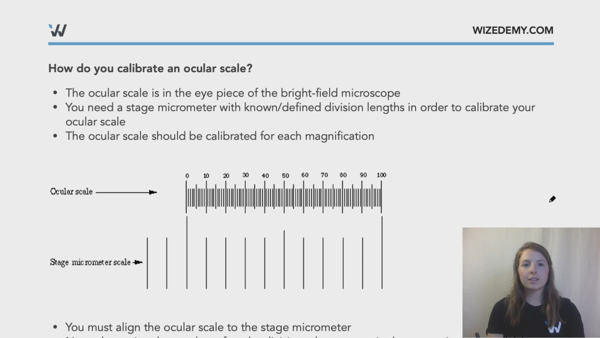
How do you calibrate an ocular scale?
- The ocular scale is in the eye piece of the bright-field microscope
- You need a stage micrometer with known/defined division lengths in order to calibrate your ocular scale
- The ocular scale should be calibrated for each magnification
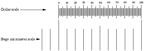
- You must align the ocular scale to the stage micrometer
- Next, determine the number of ocular divisions that span a single stage micrometer division
- In the above example 1 stage division = 10 ocular divisions
- If each stage division is 0.1 mm, then 10 ocular divisions also equals 0.1 mm
- If you need to convert between magnifications, then you need to determine the difference in magnification from the calibrated magnification to the one being used
Example: If you calibrated your microscope ocular scale using the 100x magnification and find that a single stage micrometer division spans 10 ocular divisions, what is the distance between each ocular division if a single stage micrometer division is 0.1 mm?
At 100x magnification:
= 10 um/ocular division
Converting to 40x magnification:
= 2.5
2.5 x 10 um/ocular division = 25 um/ocular division
Converting to 400x magnification:
= 0.25
0.25 x 10 um/ocular division = 2.5 um/ocular division
Converting to 10x magnification
= 10
10 x 10 um/ocular division = 100 um/ocular division
Practice: Smallest Drawing in the World
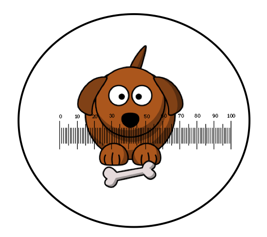
You are the judge for the world record for smallest drawing. You looking at the drawing under the microscope and using the ocular scale you need to determine the length of the drawing to see if it actually in the smallest drawing in the world. You calibrate your ocular scale using the below stage micrometer that has 0.1mm stage divisions at 100x magnification. What is the length of the below drawing if the image was captured using the 4x objective lens (eyepiece lens = 10x)?
Practice: Electron Microscopy
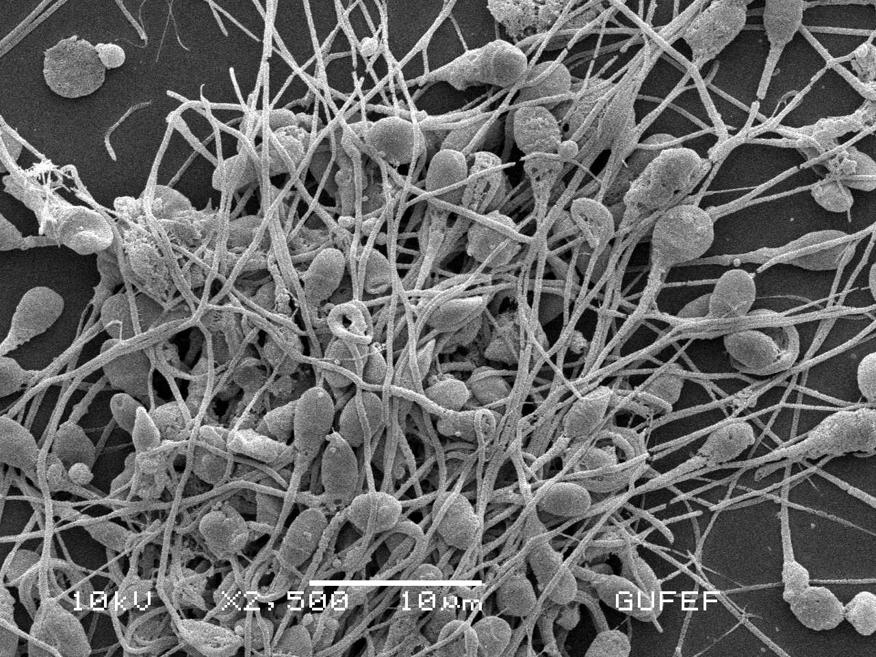
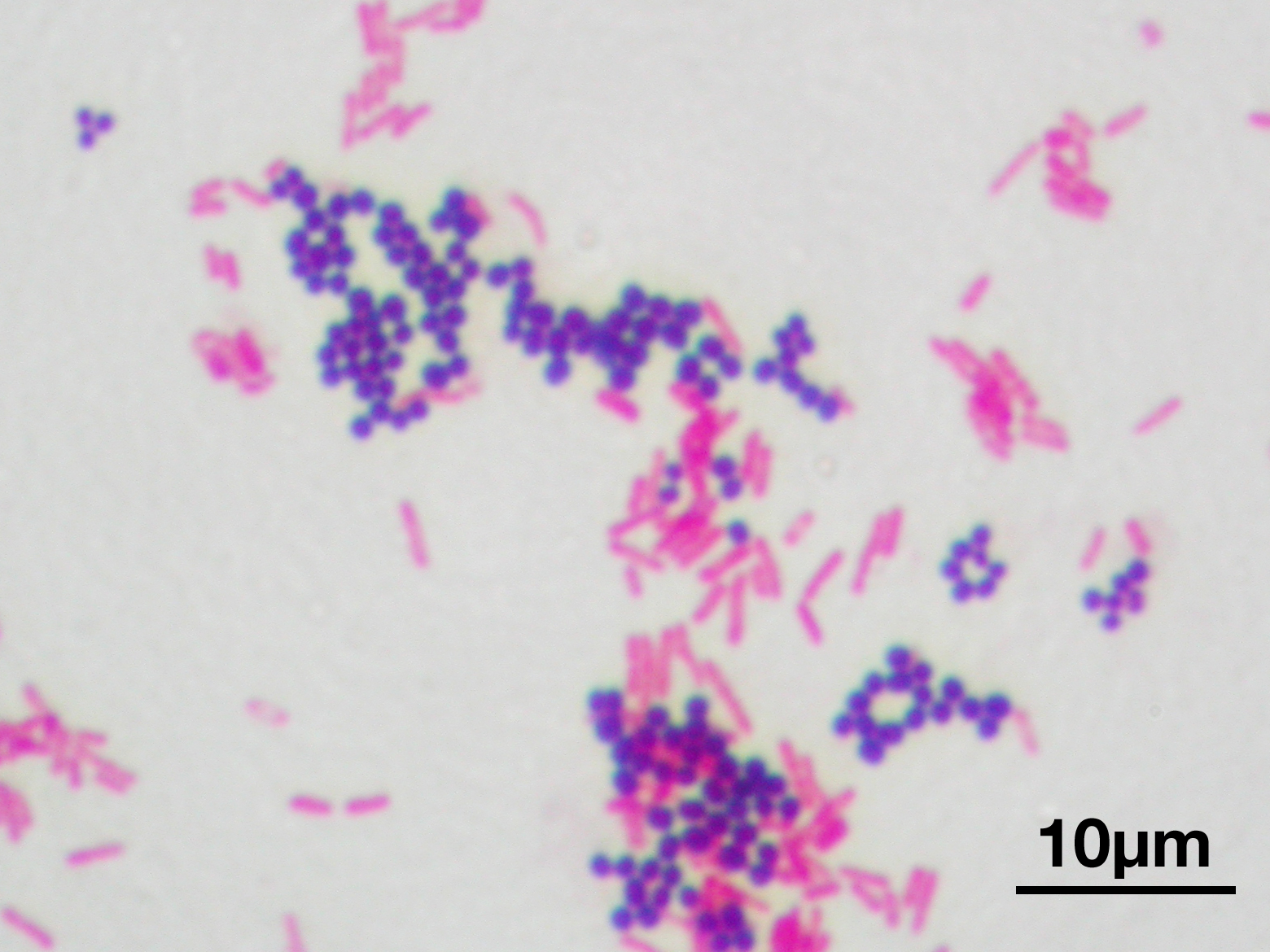
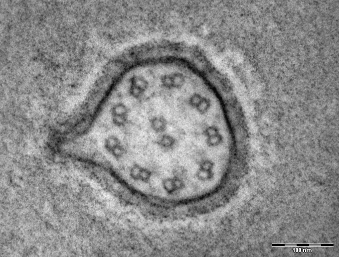
What type of microscopy was used for each of the following images?
Choose from the following options and use the three letter abbreviation:
Light microscopy (LM)
Scanning electron microscopy (SEM)
Transmission electron microscopy (TEM)
A: Human sperm cells
B: Staphylococcus and E. coli bacteria
C: Flagellum cross-section
| A | |
| B | |
| C |
Practice: Microscopy