Wize University Biochemistry Textbook > Protein Structure and Folding
Analysis of Protein Structure
0:00 / 0:00
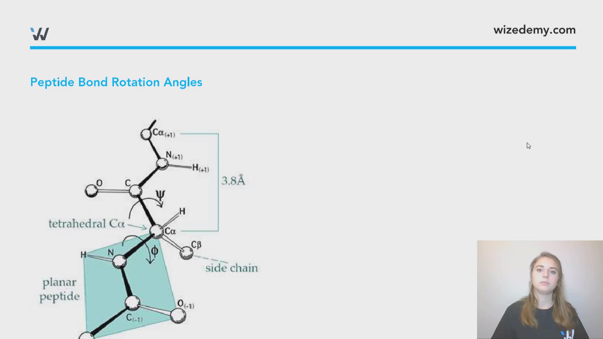
Peptide Bond Rotation Angles
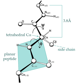
https://commons.wikimedia.org/wiki/File:PhiPsi_drawing_with_plane_and_labels.jpg. The author Dcrjsr has this file licensed under the Creative Commons Attribution 3.0 Unported license.
- The Cα - C=O and the Cα - NH bonds in the peptide backbone have measurable angles of rotations (ψ and φ respectively)
- Each secondary structure has a unique set of bond rotation angles:
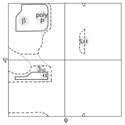
α-helix (φ,ψ) = (-60o, -50o)
β-sheet (φ,ψ) = (-140o, +135o)
Ramachandran Plots
- Ramachandran plots are created by plotting the ψ angles of a proteins amino acids on the y-axis and the φ angles on the x-axis
- This plot shows which kind of angle combinations are possible and which are not (steric hindrance)
- Since these two angels define protein geometry these plots can define protein structure
- Secondary structures show characteristic patterns on the Ramachandran plot
https://commons.wikimedia.org/wiki/File:Ramachandran_plot_original_outlines.jpg. Author Dcrjsr has image licensed under the Creative Commons Attribution 3.0 Unported license.

0:00 / 0:00
What would happen to the ramachandran plot of an alpha-helix that was denatured with urea?
Since right handed alpha-helices have a set pairing of bond angles: α-helix (φ,ψ) = (-60o, -50o), if this structure was disrupted there would be much more flexibility in the strand and those rigid angles would be more variable and less constant, drastically changing the Ramachandran plot. There would no longer be the characteristic clustering in the bottom left hand quadrant.