
0:00 / 0:00
Predicting Protein Structure
- Anfinsen's experiment shows us that secondary and tertiary structure are coded in the amino acid sequence
- Analyzing patterns in solved protein structures can lead to predictions for unknown proteins
- Ramachandran plots can be constructed through minimum energy calculations used to determine angle rotation
- These only show short-range interactions
- Tertiary predictions depend on two more factors:
- Environment: The matrix or membrane the protein exists in
- Have to take into account the small energy changes that occur through bonds breaking and forming
- Long range interactions between distant amino acids in the protein
0:00 / 0:00
Chou-Fasman Rules
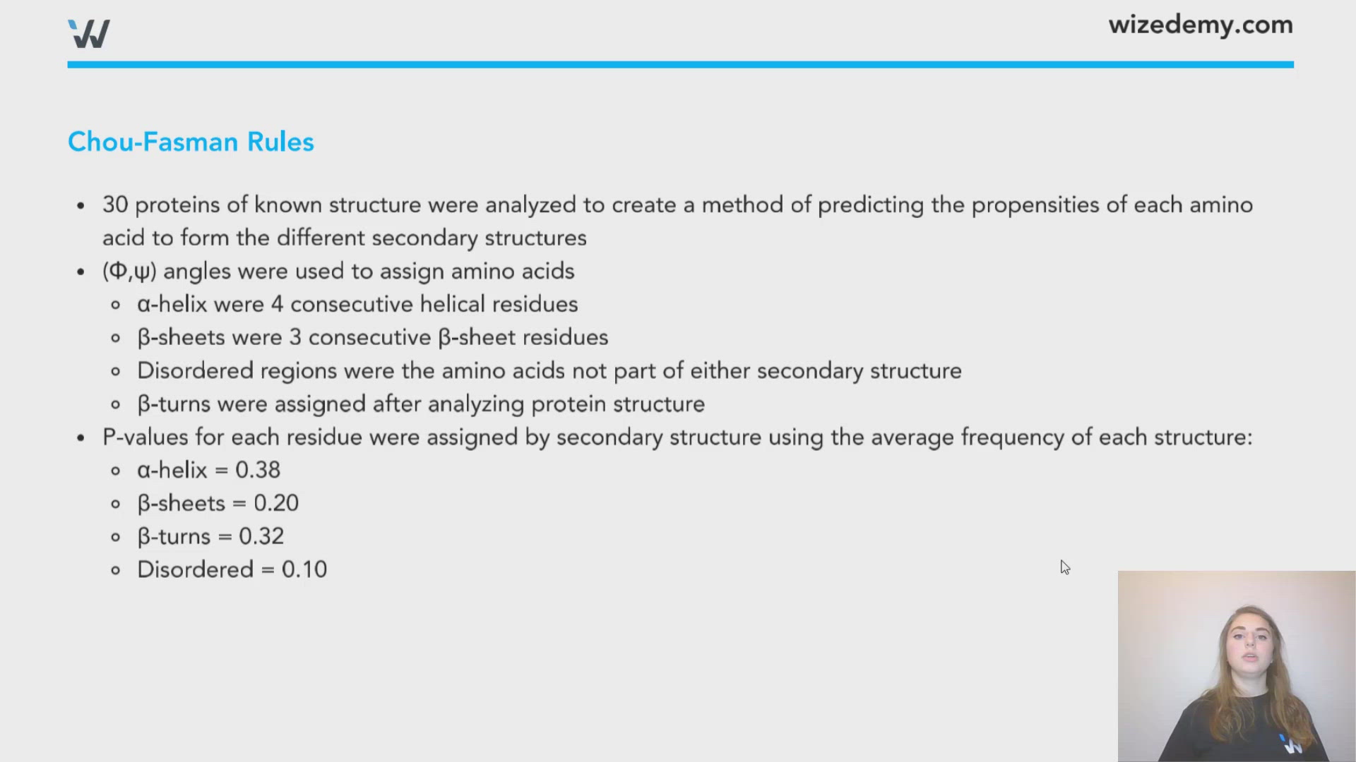
- 30 proteins of known structure were analyzed to create a method of predicting the propensities of each amino acid to form the different secondary structures
- (Φ,ψ) angles were used to assign amino acids
- α-helix were 4 consecutive helical residues
- β-sheets were 3 consecutive β-sheet residues
- Disordered regions were the amino acids not part of either secondary structure
- β-turns were assigned after analyzing protein structure
- P-values for each residue were assigned by secondary structure using the average frequency of each structure:
- α-helix = 0.38
- β-sheets = 0.20
- β-turns = 0.32
- Disordered = 0.10
- Calculation:
Pα = % AA in secondary structure/average frequency of that secondary structure
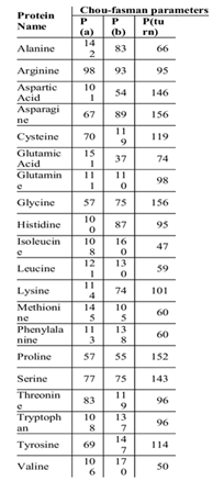
https://www.researchgate.net/figure/Chou-fasman-parameters_tbl1_301672604. Jeong et al. Available via license: Creative Commons Attribution 4.0 International.
0:00 / 0:00
Hemoglobin
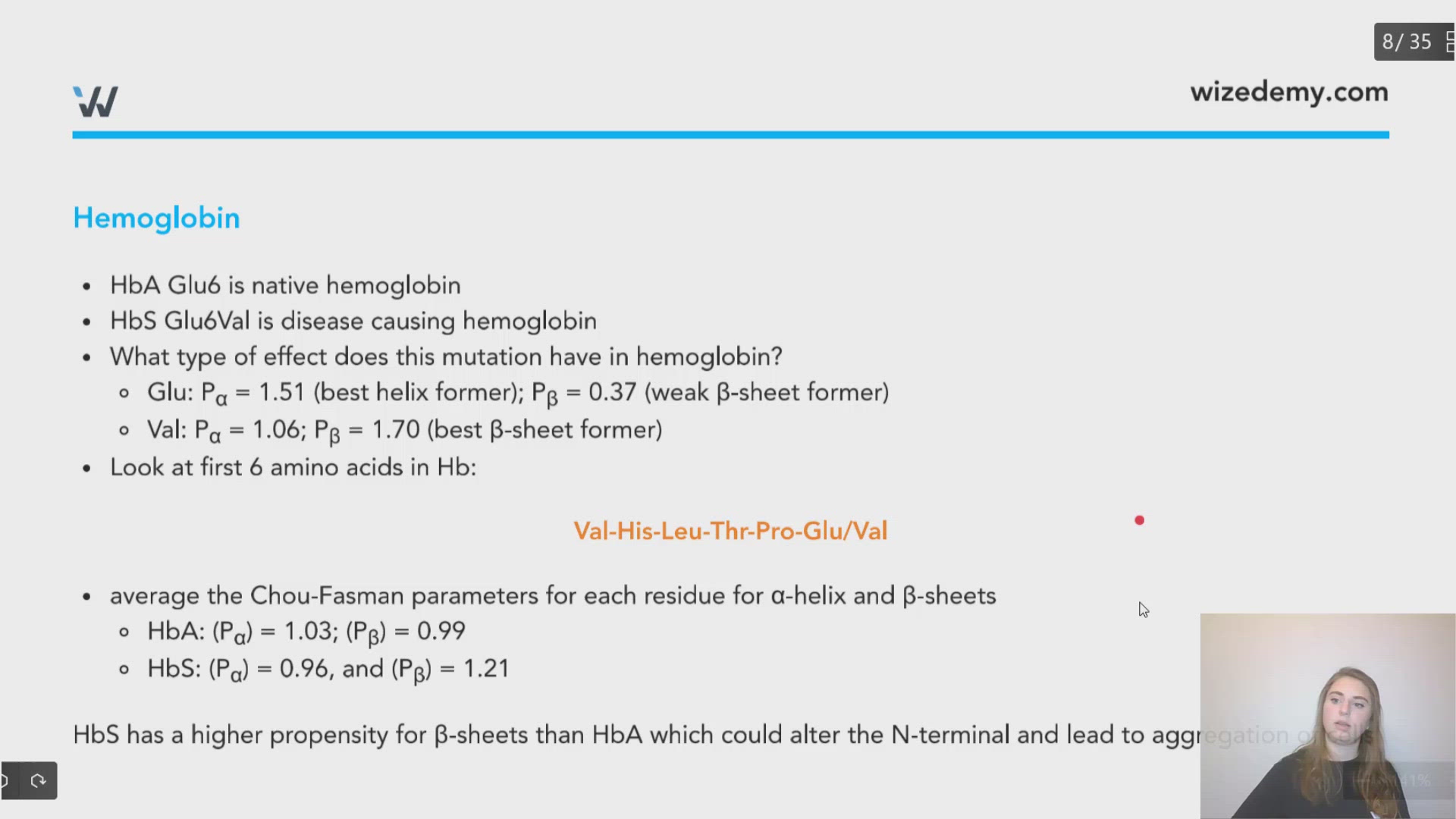
- HbA Glu6 is native hemoglobin
- HbS Glu6Val is disease causing hemoglobin
- What type of effect does this mutation have in hemoglobin?
- Glu: Pα = 1.51 (best helix former); Pβ = 0.37 (weak β-sheet former)
- Val: Pα = 1.06; Pβ = 1.70 (best β-sheet former)
- Look at first 6 amino acids in Hb:
Val-His-Leu-Thr-Pro-Glu/Val
- average the Chou-Fasman parameters for each residue for α-helix and β-sheets
- HbA: (Pα) = 1.03; (Pβ) = 0.99
- HbS: (Pα) = 0.96, and (Pβ) = 1.21
HbS has a higher propensity for β-sheets than HbA which could alter the N-terminal and lead to aggregation of cells
Glucagon
- Depending on concentration the C-terminal will either be predominantly helical or predominantly β-sheets
- As above, determine the C-terminal's propensity to form α-helix and β-sheets:
Ala-Gln-Asp-Phe-Val-Gln-Trp-Leu-Met-Asn-Thr
- HbA: (Pα) = 1.18; (Pβ) = 1.15
- These are very similar values, indicating glucagon has equal an propensity to form either secondary structure
Exam Tip
If a questions asks you: what secondary structure will most likely be formed by a given set of amino acids? First determine the average Chou-Fasman parameters for each secondary structure by adding all of the individual parameters then dividing by the number of participating amino acids. After that compare values. The highest value indicates the preferred secondary structure.

0:00 / 0:00
Why can't Ramachandran plots predict tertiary structure?
Ramachandran plots measure angles of bond rotation in amino acid backbones. Since secondary structures have consistent and recurring features, they create patterns in the plot specific to each secondary structure. Tertiary structure on the other hand depend on additional factors such as the environment of the protein and long-range interactions between amino acids. These more "random" interactions will not provide useful information on a Ramachandran plots as there are no recurring patterns that involve several amino acids holding the same spacial conformations.
0:00 / 0:00
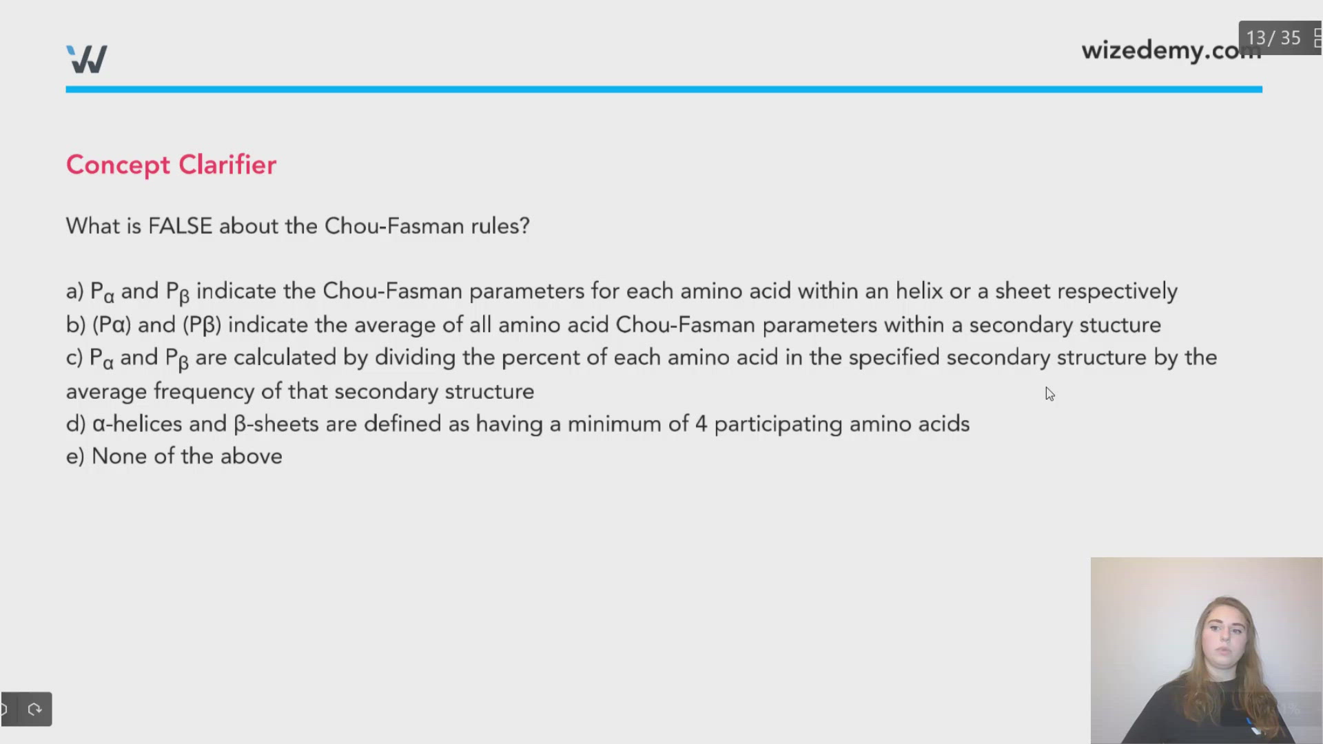
What is FALSE about the Chou-Fasman rules?
a) Pα and Pβ indicate the Chou-Fasman parameters for each amino acid within an helix or a sheet respectively
b) (Pα) and (Pβ) indicate the average of all amino acid Chou-Fasman parameters within a secondary structure
c) Pα and Pβ are calculated by dividing the percent of each amino acid in the specified secondary structure by the average frequency of that secondary structure
d) α-helices and β-sheets are defined as having a minimum of 4 participating amino acids
e) None of the above
d) α-helices and β-sheets are defined as having a minimum of 4 participating amino acids. This is false. Sheets are defined as a minimum of 3.
Using Chou-Fasman parameters:
Pα for Leu = 1.21; Pα for Glu = 1.51; Pα for Val = 1.06
Pβ for Leu = 1.30; Pβ for Glu = 0.37; Pβ for Val = 1.70
What structure would you predict the following peptide to have: VVLEVLVVL